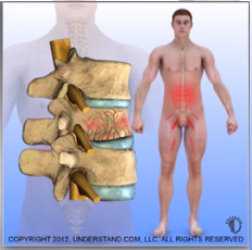
Vertebral compression fractures are associated with significant performance impairments in physical, functional, and psychosocial domains particularly in older women.[1]However, medical and surgical options are now available that can relieve the severe pain and disability from these fractures. Fractures in the lumbar spine occur for a number of reasons. In younger patients, fractures are usually due to violent trauma. Car accidents frequently cause flexion and flexion distraction injuries. Jumps or falls from heights cause burst fractures. These fractures can also result in serious neurological injury. In older patients, lumbar compression fractures usually occur in the absence of trauma, or in the context of minor trauma, such as a fall. The most common underlying reason for these fractures in geriatric patients, especially women, is osteoporosis or pathologic weakening of the bone. Other disorders that can contribute to the occurrence of compression fractures include malignancy, infections, and renal disease.
Different types of fractures can occur in the lumbar (or thoracic) spine. Classification of these fractures is based on the 3-column anatomic theory of Denis, which describes anterior, middle, and posterior spinal columns consisting of aspects of the spine and their corresponding ligaments and other soft-tissue elements. The Denis system, however, was created to classify traumatic fractures. A similar classification system does not exist for compression fractures. The main reason to use such a classification is to help determine whether a fracture is stable. Instability in the Denis system implies that damage has occurred to at least 2 of the columns of the lumbar spine.
Wedge fractures are the most common type of lumbar fracture and are the typical compression fracture of malignancy or osteoporosis. They occur as a result of an axially directed central compressive force as.
Fractures involving flexion and distraction forces are often due to lap belts in motor vehicle accidents. Commonly, the posterior columns are compromised in these injuries because the ligaments of the posterior elements are disrupted. This type of injury is quite common in young children. Most patients with flexion-distraction injuries remain neurologically intact.
• Burst fractures result from high-energy axial loads to the spine. Multiple classification systems exist for these fractures. The severity of the deformity, the severity of canal compromise, the extent of loss of vertebral body height, and the degree of neurologic deficit affect the determination of whether these injuries are unstable.
When any of the above injuries occurs with a severe rotational force, the degree of injury and of instability increases.
In osteoporosis, osteoclastic activity exceeds osteoblastic activity, resulting in a generalized decrease in bone density. The osteoporosis weakens the bone to the point that even a minor fall on the tailbone, causing an axial load or flexion, results in one or more compression fractures. The fracture is usually wedge shaped. Without correction, a wedge fracture invariably increases the degree of kyphosis.
Midline back pain is the hallmark symptom of lumbar compression fractures. The pain is axial, nonradiating, aching, or stabbing in quality and may be severe and disabling. The location of the pain corresponds to the fracture site, as seen on radiographs. In elderly patients with severe osteroporosis, however, there may be no pain at all as the fracture occurs spontaneously.
Vertebral fractures may also cause referred pain. Gibson et al presented a study of 350 patient encounters in 288 patients with 1 or more compression fracture without spinal nerve compression. They found that nonmidline pain was present in 240 of the 350 encounters. The pain was typically in the ribs, hip, groin, or buttocks. Treatment of the fracture with vertebroplasty (see Other Treatment) resulted in 83% of those patients gaining pain relief.
Alternatively, many compression fractures are painless. Osteoporosis is a silently progressive disease. Osteoporotic compression fractures are often diagnosed when an elderly patient presents with symptoms such as progressive scoliosis or mechanical lower back pain and the physician obtains routine lumbar radiographs.
Surgical intervention is required when neurologic dysfunction and/or instability occurs as a result of the lumbar fracture.
Neurologic problems may manifest in many ways. Reduced leg strength (paresis) or complete weakness (paralysis) is an obvious problem. Loss of sensation in the lower extremities and in the perianal area (saddle anesthesia) can be just as important. Urinary retention and urinary and fecal incontinence are very important signs that indicate the need for surgery.
The extent of neurologic problems also depends on whether there is compression of the lower portion of the spinal cord (conus medullaris) or lumbar nerve roots (cauda equina).
Clinical instability is manifested primarily by severe pain that does not improve or that worsens with time. Patients with clinical instability may require surgery. This information is corroborated radiographically by visualizing kyphotic deformity on plain radiographs and disruption of the interspinous ligaments on MRIs.
Radiographic instability refers to cases in which the ligamentous disruption is severe, canal compromise occurs to a degree that neurologic symptoms are present, and movement of the fracture fragments is seen on dynamic or motion radiographs. These fractures almost always need surgical fixation, although on rare occasions a rigid brace will suffice. Some patients who initially are braced may show gradual worsening of symptoms on radiographs, with findings of progressive kyphosis with loss of vertebral body height. These patients also require surgical intervention.
The surgical procedure used for correction of a lumbar fracture depends on certain factors. These critical factors include the degree of bony canal compromise seen, the level of fracture, neurologic examination findings, the curvature of the spine and the patient’s health status.
Verterbral Augmentation or Kyphoplasty is a minimally invasive procedure utilized to reduce pain and restore vertebral bony support.
In the setting of spinal deformity or spinal nerve impingement a minimally invasive decompression and/or stabilization procedure may be required. The choice of an anterior or a posterior approach for decompression is dictated by the location and severity of bony canal compromise.
Once a patient has undergone surgery, a brace is prescribed in the postoperative period, depending on the etiology of the fracture.
The time period for healing from surgery is variable for individual patients, depending on their health status.

Dr. Ram R. Vasudevan, MD, FAANS
Austin NeuroSpine PLLC
5300 Bee Cave Road, Building 1,
Suite 220 Austin, TX 78746
Phone: (512) 640-0010
Monday: 8:00 AM – 5:00 PM
Tuesday: 8:00 AM – 5:00 PM
Wednesday: 8:00 AM – 5:00 PM
Thursday: 8:00 AM – 5:00 PM
Friday: 8:00 AM – 5:00 PM
Saturday: Closed
Sunday: Closed
© 2023 Austin NeuroSpine PLLC. All rights reserved.