The spinal column is one of the most vital parts of the human body, supporting our trunks and making all of our movements possible. Its anatomy is extremely well designed, and serves many functions, including:
All of the elements of the spinal column and vertebrae serve the purpose of protecting the spinal cord, which provides communication to the brain and mobility and sensation in the body through the complex interaction of bones, ligaments and muscle structures of the back and the nerves that surround it.
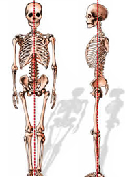
The normal adult spine is balanced over the pelvis, requiring minimal workload on the muscles to maintain an upright posture.
Loss of spinal balance can result in strain to the spinal muscles and spinal deformity. When the spine is injured and its function impaired, the consequences may be painful and even disabling.
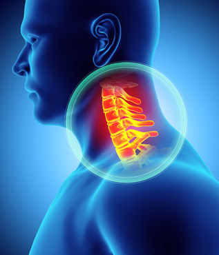
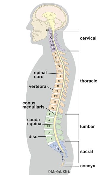
Humans are born with 33 separate vertebrae. By adulthood, we typically have 24 due to the fusion of the vertebrae in the sacrum.
All of the elements of the spinal column and vertebrae serve the purpose of protecting the spinal cord, which provides communication to the brain and mobility and sensation in the body through the complex interaction of bones, ligaments and muscle structures of the back and the nerves that surround it.
The normal adult spine is balanced over the pelvis, requiring minimal workload on the muscles to maintain an upright posture.
Loss of spinal balance can result in strain to the spinal muscles and spinal deformity. When the spine is injured and its function impaired, the consequences may be painful and even disabling.
The elements of the spine are designed to protect the spinal cord, support the body and facilitate movement.
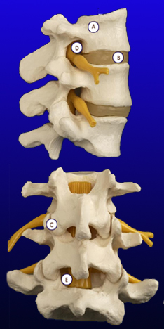
A. Vertebrae
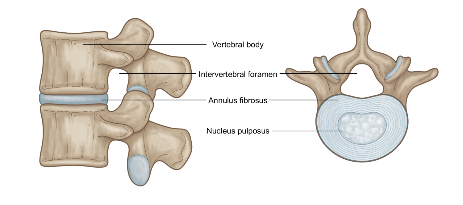
The vertebrae support the majority of the weight imposed on the spine. The body of each vertebra is attached to a bony ring consisting of several parts. A bony projection on either side of the vertebral body called the pedicle supports the arch that protects the spinal canal. The laminae are the parts of the vertebrae that form the back of the bony arch that surrounds and covers the spinal canal. There is a transverse process on either side of the arch where some of the muscles of the spinal column attach to the vertebrae. The spinous process is the bony portion of the vertebral body that can be felt as a series of bumps in the center of a person’s neck and back.
B. Intervertebral Disc
Between the spinal vertebrae are discs, which function as shock absorbers and joints. They are designed to absorb the stresses carried by the spine while allowing the vertebral bodies to move with respect to each other. Each disc consists of a strong outer ring of fibers called the annulus fibrosis, and a soft center called the nucleus pulposus. The outer layer (annulus) helps keep the disc’s inner core (nucleus) intact. The annulus is made up of very strong fibers that connect each vertebra together. The nucleus of the disc has a very high water content, which helps maintain its flexibility and shock-absorbing properties.
C. Facet Joint
The facet joints connect the bony arches of each of the vertebral bodies. There are two facet joints between each pair of vertebrae, one on each side. Facet joints connect each vertebra with those directly above and below it, and are designed to allow the vertebral bodies to rotate with respect to each other.
D. Neural Foramen
The neural foramen is the opening through which the nerve roots exit the spine and travel to the rest of the body. There are two neural foramen located between each pair of vertebrae, one on each side. The foramen creates a protective passageway for the nerves that carry signals between the spinal cord and the rest of the body.
E. Spinal Cord and Nerves
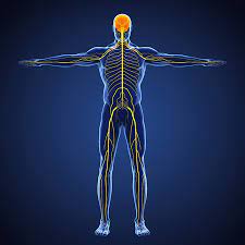
The spinal cord extends from the base of the brain to the area between the bottom of the first lumbar vertebra and the top of the second lumbar vertebra. The spinal cord ends by diverging into individual nerves that travel out to the lower body and the legs. Because of its appearance, this group of nerves is called the cauda equina – the Latin name for “horse’s tail.” The nerve groups travel through the spinal canal for a short distance before they exit the neural foramen.
The spinal cord is covered by a protective membrane called the dura mater, which forms a watertight sac around the spinal cord and nerves. Inside this sac is spinal fluid, which surrounds the spinal cord.
The nerves in each area of the spinal cord are connected to specific parts of the body. Those in the cervical spine, for example, extend to the upper chest and arms; those in the lumbar spine the hips, buttocks and legs. The nerves also carry electrical signals back to the brain, creating sensations. Damage to the nerves, nerve roots or spinal cord may result in symptoms such as pain, tingling, numbness and weakness, both in and around the damaged area and in the extremities.
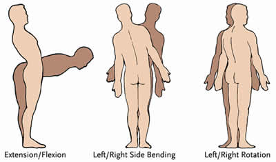
Many muscle groups that move the trunk and the limbs also attach to the spinal column. The muscles that closely surround the bones of the spine are important for maintaining posture and helping the spine to carry the loads created during normal activity, work and play. Strengthening these muscles can be an important part of physical therapy and rehabilitation.
All of the elements of the spinal column and vertebrae serve the purpose of protecting the spinal cord, which provides communication to the brain, mobility and sensation in the body through the complex interaction of bones, ligaments and muscle structures of the back and the nerves that surround it.
The true spinal cord ends at approximately the L1 level, where it divides into the many different nerve roots that travel to the lower body and legs. This collection of nerve roots is called the cauda equina, which means “horse’s tail,” and describes the continuation of the nerve roots at the end of the spinal cord.
Unless Noted Otherwise, All Articles and Graphics Copyright ©2013, Medtronic Sofamor Danek, All Rights Reserved.
Please review our Privacy Policy, Editorial Policy, or Terms Of Use for more information.

Dr. Ram R. Vasudevan, MD, FAANS
Austin NeuroSpine PLLC
5300 Bee Cave Road, Building 1,
Suite 220 Austin, TX 78746
Phone: (512) 640-0010
Monday: 8:00 AM – 5:00 PM
Tuesday: 8:00 AM – 5:00 PM
Wednesday: 8:00 AM – 5:00 PM
Thursday: 8:00 AM – 5:00 PM
Friday: 8:00 AM – 5:00 PM
Saturday: Closed
Sunday: Closed
© 2023 Austin NeuroSpine PLLC. All rights reserved.